
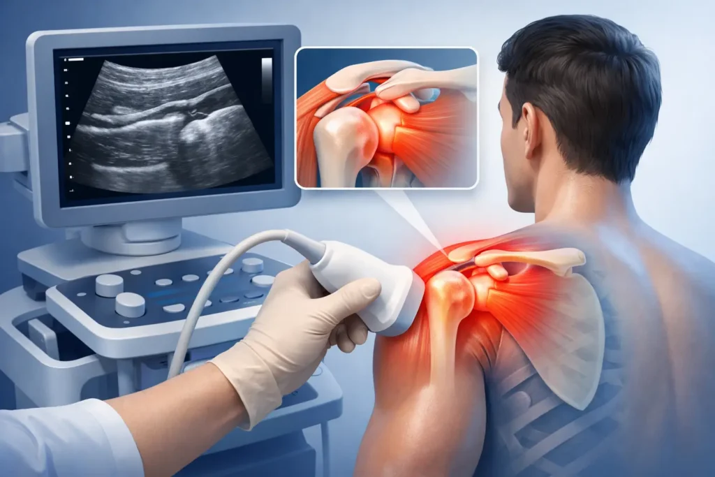
A shoulder that hurts when you reach for a seatbelt, lift a kettle or lie on one side can quickly affect work, sleep and exercise. Ultrasound diagnostics for shoulder pain can provide fast, accurate insight into the structures that may be causing those symptoms, often during the same clinical appointment.
Shoulder pain is not one condition. It can arise from a tendon, bursa, joint, muscle, ligament or referred pain from the neck. A detailed assessment remains essential, but diagnostic ultrasound allows a specialist to look beneath the surface, compare both shoulders and see how key structures behave as you move. That clarity helps build the best treatment plan sooner, rather than relying on guesswork or treating a scan report in isolation.
What an ultrasound scan can show in a painful shoulder
Ultrasound uses high-frequency sound waves to create real-time images. There is no radiation, no injection of contrast dye and, for most people, no special preparation. A clinician places a small probe against the skin with gel and examines the shoulder from different angles.
The scan is particularly useful for the soft tissues around the shoulder. This includes the rotator cuff tendons, the long head of biceps tendon, the subacromial-subdeltoid bursa, parts of the joint capsule and the acromioclavicular joint at the top of the shoulder. It may identify tendon thickening, inflammation, calcific deposits, fluid around a tendon, bursitis, partial or full-thickness tendon tears, biceps instability and some joint changes.
One of ultrasound’s major advantages is that it is dynamic. Rather than viewing a shoulder only while it is still, the clinician can ask you to lift or rotate your arm during the examination. This can help assess painful movement patterns, tendon movement and impingement-related symptoms in real time. Comparing the painful side with the other shoulder can also put minor changes into useful context.
Why the diagnosis matters before treatment
Shoulder pain is often labelled broadly as a strain or impingement. Sometimes that is enough to begin sensible rehabilitation. But if pain is persistent, marked, follows an injury or has not improved with initial treatment, a more precise diagnosis can change the direction of care.
For example, a painful arc when lifting the arm may be linked to rotator cuff tendinopathy, bursal irritation, a calcific deposit or more than one problem at once. Each can require a different emphasis in rehabilitation. Tendon-related pain may benefit from a progressive loading programme, while significant bursitis may require pain management to make movement and exercise tolerable. A substantial tear after a fall may warrant a different referral pathway altogether.
At FAB Clinic, ultrasound findings are considered alongside your history, physical examination, strength, range of movement, work demands and goals. A scan is valuable because it informs clinical judgement. It is not a standalone verdict on whether you should be in pain.
When ultrasound diagnostics for shoulder pain are useful
An ultrasound assessment can be particularly helpful when symptoms are limiting daily activity, sleep or sport, or when progress has stalled. It is commonly considered after a lifting injury, fall, sudden traction injury or a gradual onset of pain that has become more persistent.
It can also be useful if you have weakness when lifting the arm, pain at night, clicking or catching, pain over the front or outer shoulder, or tenderness at the top of the shoulder. For active people, the ability to assess a painful shoulder during movement can be especially relevant when symptoms occur only with a particular gym exercise, tennis serve or swimming stroke.
Not every shoulder problem needs immediate imaging. Many uncomplicated episodes improve with an expert assessment, activity modification and a structured rehabilitation plan. Equally, an ultrasound scan should not be delayed when the clinical picture suggests that a tendon injury, marked inflammation or another significant soft-tissue problem needs clarification.
What happens during your appointment
Your appointment should begin with questions that give the scan meaning. Your clinician will ask how the pain started, where it is felt, which movements provoke it, whether there has been a specific injury and how symptoms affect sleep, work and exercise. They will then examine posture, movement, strength and relevant areas such as the neck and upper back.
During the scan, you will usually be seated and asked to expose the shoulder area. The clinician applies gel and moves the probe over several regions of the shoulder. You may be asked to position your arm in different ways or perform controlled movements. The examination is usually comfortable, although pressing directly over an already sore area can be tender.
A key benefit of specialist ultrasound is immediate discussion. Your clinician can explain what is visible, what may be relevant to your symptoms and what remains uncertain. You should leave with a clear understanding of the recommended next step, whether that is rehabilitation, hands-on treatment, shockwave therapy for appropriate tendon conditions, a guided injection consultation or further imaging or referral.
Ultrasound versus MRI and X-ray
Ultrasound is excellent for many tendon, bursa and superficial soft-tissue conditions. It is quick, accessible and dynamic, making it a strong first-line imaging choice for many shoulder presentations.
However, it has limits. MRI may be more appropriate where there is concern about deeper joint structures, cartilage, the labrum, bone marrow changes or complex surgical planning. An X-ray can be helpful when arthritis, fracture, bone shape changes or significant calcification are suspected. In some cases, a clinician may recommend more than one test because each answers a different question.
The right investigation depends on your symptoms and examination findings. The aim is not to order every possible scan. It is to use the test most likely to give meaningful information and guide the next stage of recovery.
From scan findings to a treatment plan
Finding a structural change does not automatically mean it is the source of pain. Tendon changes and small tears can exist without symptoms, particularly as we get older. Conversely, a shoulder can be very painful even when imaging changes are relatively modest. This is why expert interpretation matters.
If the findings support rotator cuff tendinopathy, treatment may focus on managing aggravating activities, restoring movement and gradually rebuilding shoulder strength and control. For bursitis, reducing irritation while maintaining gentle movement may be the priority. Calcific tendinopathy can need a more targeted approach, particularly when pain is severe or persistent.
Where clinically appropriate, ultrasound guidance can also improve the accuracy of procedures such as a joint or bursal injection. The clinician can see the needle pathway and target area in real time. This does not replace rehabilitation. Pain relief can create the opportunity to move better and engage fully with a progressive recovery programme, which is often central to longer-term improvement.
When shoulder pain needs urgent assessment
Seek urgent medical attention if shoulder pain follows a major injury, the shoulder looks deformed, you cannot move the arm after trauma, or there is sudden severe weakness. Prompt assessment is also needed if the shoulder is hot, red and swollen, especially with fever or feeling unwell.
Pain spreading into the chest, jaw or arm, breathlessness, sweating or nausea needs emergency medical attention, as these symptoms may not be caused by the shoulder itself. Persistent unexplained pain, significant weight loss or new neurological symptoms should also be assessed promptly.
A good shoulder assessment gives you more than a name for the problem. It gives you a practical route back to comfortable movement, stronger function and confidence in what your shoulder can do next.