
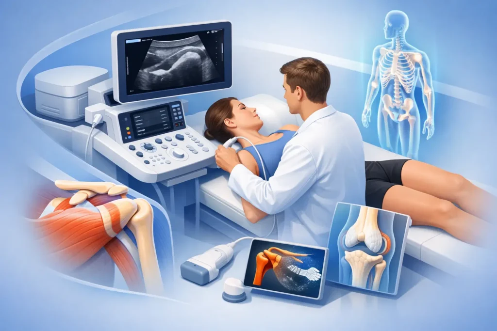
A painful shoulder that catches when you reach overhead, a swollen knee after a run, or heel pain that has lingered for months can all leave one question unanswered: what is actually causing it? A guide to musculoskeletal ultrasound scans can help explain why this imaging tool is so valuable when symptoms need fast, accurate insight.
Musculoskeletal ultrasound uses sound waves to create live images of structures close to the skin. It allows a specialist to assess muscles, tendons, ligaments, joints, bursae and other soft tissues while you are in the clinic. Unlike a static image alone, ultrasound can often show how a structure behaves during movement, helping connect what appears on screen with the pain or restriction you feel day to day.
What is a musculoskeletal ultrasound scan?
A musculoskeletal, or MSK, ultrasound scan is a non-invasive diagnostic examination. A clinician applies clear gel to the area being assessed and moves a small handheld probe over the skin. The probe sends and receives high-frequency sound waves, producing images on a screen in real time.
There is no radiation involved, no need for injections for a standard diagnostic scan, and most appointments are straightforward. The scan is usually comfortable, although pressure over a particularly inflamed or injured area may reproduce familiar symptoms. That response can itself provide useful clinical information.
At FAB Clinic, ultrasound is used as part of an integrated assessment rather than as an isolated test. Your symptoms, medical history, physical examination and scan findings are considered together to build the best treatment plan. This matters because an image may show a change in a tendon or joint that is not necessarily the source of pain, while a clinically significant problem may be clearer when the scan is performed dynamically.
What can musculoskeletal ultrasound scans show?
Ultrasound is particularly effective for many common soft-tissue and joint concerns. It can identify inflammation, fluid, tissue thickening, tears and changes associated with wear, overload or injury. It is frequently used to assess the shoulder, elbow, wrist, hand, hip, knee, ankle and foot.
For example, a shoulder scan may help assess the rotator cuff tendons, bursa and biceps tendon in someone with painful lifting or night pain. At the elbow, ultrasound can support diagnosis of tendon changes linked with tennis elbow or golfer’s elbow. In the foot, it is often helpful for plantar fasciitis, Achilles tendon pain, ankle ligament injuries and forefoot problems.
A scan may show tendon thickening or degeneration, a partial tear, bursitis, joint fluid, synovial inflammation, a ganglion cyst, calcification or a muscle injury. It can also assist with the assessment of superficial nerves and soft-tissue lumps. In some cases, comparing the painful side with the unaffected side offers a clearer picture.
The value of dynamic imaging
One of ultrasound’s strongest advantages is that it can be used while you move. A clinician may ask you to lift your arm, bend your knee, rotate your ankle or grip with your hand as they watch relevant tissues in action.
This can be helpful where pain occurs only in a particular position or activity. In the shoulder, for instance, dynamic assessment may reveal whether structures are being irritated during certain movements. It gives the practitioner a practical view of function, not just anatomy.
What ultrasound cannot tell us on its own
Ultrasound is an excellent tool, but it is not the answer to every musculoskeletal problem. Sound waves do not pass well through bone, so ultrasound is less suitable for viewing structures deep within a joint or assessing many bone injuries. It also has limitations for some spinal conditions and deep pelvic or hip structures.
If a fracture, significant bone change, complex cartilage injury, deep ligament injury or nerve problem is suspected, an X-ray, MRI, CT scan, blood tests or specialist referral may be more appropriate. The right investigation depends on your symptoms, examination findings and clinical history. Good care is not about ordering the most tests – it is about choosing the investigation most likely to change your treatment.
When might you need an MSK ultrasound scan?
A scan may be recommended when pain has not improved as expected, when there is swelling or weakness, or when the diagnosis remains unclear after an initial assessment. It is also useful where a clinician needs to distinguish between several possible causes of similar symptoms.
You may benefit from musculoskeletal ultrasound if you have persistent shoulder pain, a suspected tendon or muscle tear, recurrent joint swelling, a sports injury, heel or Achilles pain, wrist or thumb pain, or a lump around a joint. It may also be considered before an ultrasound-guided injection, where accurate needle placement is essential.
However, a scan is not always needed immediately after an injury. Many straightforward strains, mild sprains and short-lived episodes of back or neck pain can be managed safely with an expert assessment and a structured rehabilitation plan. Early imaging can occasionally find incidental changes that sound worrying but are not causing symptoms. Your clinician should explain why a scan is likely to be useful in your individual case.
What happens during your appointment?
Most ultrasound assessments begin with a focused conversation. Your clinician will ask how the problem started, which movements trigger it, whether pain is constant or intermittent, and how it affects work, sport, sleep and daily activity. They will also check for relevant medical history, previous injuries and warning signs that may require a different pathway.
You may need to expose the area being examined, such as the shoulder, knee or foot, but your comfort and privacy should be respected throughout. Gel is then applied to the skin. It can feel cool, but it washes away easily and does not stain clothing.
The scanning portion is commonly completed within 15 to 30 minutes, though timing varies according to the body area and complexity of the issue. You may be asked to move, stand, sit or lie in different positions. If the scan identifies a tender area, the clinician may gently press with the probe to see whether it reproduces your usual pain.
The real benefit of an in-clinic scan is the opportunity to discuss the findings at the same appointment. Rather than waiting for an image report without context, you can understand what has been seen, what it may mean and what happens next.
Preparing for a musculoskeletal ultrasound scan
For most MSK scans, no special preparation is required. Wear loose, comfortable clothing that gives easy access to the body part being assessed. Avoid applying thick creams or oils to the skin on the day if possible, as these can make the probe harder to position.
Bring details of relevant previous scans, X-rays, hospital letters and medication if you have them. If you are taking blood-thinning medication or have a medical condition that may affect a planned injection, tell your clinician in advance. This does not usually prevent a diagnostic scan, but it may affect treatment decisions.
You can normally eat, drink, drive and return to work or normal activities straight after a diagnostic ultrasound. If an injection procedure is planned, you will receive specific advice about preparation and aftercare.
From scan findings to a recovery plan
A scan is valuable when it leads to a better decision. Depending on the findings, your plan might include targeted physiotherapy, activity modification, hands-on treatment, progressive strengthening, shockwave therapy, pain-management strategies or onward referral. The aim is not simply to name a tissue problem, but to reduce pain, restore confidence in movement and improve function.
Where clinically appropriate, ultrasound can also guide joint, tendon sheath or bursa injections. Visualising the needle in real time helps the practitioner place treatment accurately while avoiding nearby structures. An injection is not automatically the right answer, and it works best when used for a clear indication alongside a rehabilitation plan rather than as a substitute for one.
A finding such as tendinopathy does not always mean you need to stop all activity. Often, the best approach is to modify load temporarily and rebuild tolerance gradually. Conversely, a significant tear, marked weakness or signs of infection may need more urgent medical assessment. Your treatment should reflect the whole clinical picture, not a scan result in isolation.
Questions worth asking after your scan
Before you leave, make sure you understand what the scan has shown and how confident the clinician is that it explains your symptoms. Ask what you can continue doing, which activities to adjust, whether further tests are needed, and what improvement should look like over the next few weeks.
Clear answers turn imaging into action. If pain is limiting the way you work, train, sleep or move, a timely musculoskeletal ultrasound assessment can provide the clarity needed to move forward with confidence and the right support.