
That nagging tendon pain often starts as something easy to dismiss – a sore Achilles after a run, a sharp pull in the shoulder when reaching overhead, or an elbow that complains every time you lift a bag. The problem is that tendon symptoms can look similar on the surface while needing very different treatment underneath. That is where diagnostic ultrasound for tendon pain becomes so valuable. It gives fast, accurate insight into what the tendon is doing in real time, helping shape a treatment plan that is based on findings rather than guesswork.
For many patients, the frustrating part is not just the pain itself. It is the uncertainty. You may have rested, stretched, iced the area, tried exercises from the internet, or even had standard physiotherapy elsewhere, yet the symptoms keep returning. Tendons do not always settle quickly, and they do not all fail in the same way. Some are overloaded and irritated. Some are degenerative. Some have small tears. Some are reacting because another structure nearby is driving the problem. Good treatment starts with identifying which of those situations you are dealing with.
Why diagnostic ultrasound for tendon pain is so useful
Diagnostic ultrasound is particularly effective in musculoskeletal care because it allows a clinician to assess soft tissues dynamically. In plain terms, that means the tendon can be seen while it moves, contracts, glides and responds to pressure. This matters because tendon pain is not always obvious when the body is still.
An X-ray will not show a tendon properly. An MRI can be very useful in some cases, but it is not always the first or fastest option, and it does not give the same live, movement-based assessment. Ultrasound offers a practical middle ground – detailed enough to identify many common tendon problems, quick enough to support immediate clinical decisions, and efficient enough to integrate directly into a wider treatment pathway.
For patients, the benefit is straightforward. Instead of being told to wait and see, you can often get a clearer answer at the point of assessment. That can shorten the time between symptoms, diagnosis and targeted treatment.
What ultrasound can show in a painful tendon
A healthy tendon has a clear, organised fibre pattern. When pain develops, ultrasound can reveal changes in that structure. These may include thickening of the tendon, disrupted fibres, local swelling, fluid around the tendon, calcification, neovascularity linked with chronic tendon change, or partial tearing.
That information helps distinguish between different stages and types of tendon injury. For example, an overloaded patellar tendon in an active gym-goer may need a very different rehabilitation approach from a chronic rotator cuff tendon that has been degenerating over time. Both may hurt, but the management is not interchangeable.
Ultrasound can also assess structures around the tendon. Sometimes the real issue is not just within the tendon itself but in the sheath, the bursa, or an adjacent joint. Shoulder pain is a good example. Patients often assume they have a tendon problem, but imaging may show bursitis, impingement, calcific change, or a combination of issues. That level of detail allows treatment to become more precise.
Common tendon problems where ultrasound helps
Diagnostic ultrasound is regularly used for tendon pain affecting the shoulder, elbow, wrist, hip, knee, ankle and foot. In practice, that often means rotator cuff pain, tennis elbow, gluteal tendinopathy, patellar tendinopathy, Achilles tendinopathy and plantar fascia-related heel pain where tendon and soft tissue overload can overlap.
It is especially helpful when symptoms have not improved as expected, when the diagnosis is unclear, or when there is concern about a tear. It can also be valuable for athletes or active adults who need to know whether it is safe to continue training, modify activity, or stop altogether for a period.
That said, ultrasound is not about chasing scans for every minor ache. A specialist assessment still comes first. Imaging is most useful when it changes decision-making, confirms suspected findings, or helps rule out a more significant problem.
What happens during a diagnostic ultrasound appointment
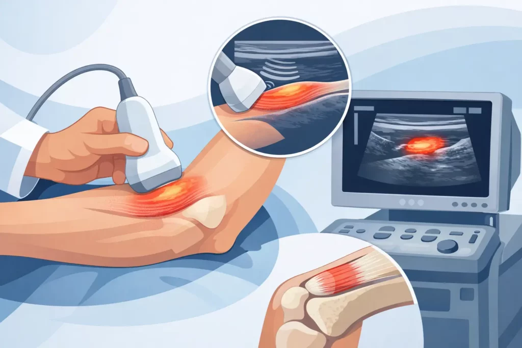
The scan itself is straightforward and generally very well tolerated. Gel is applied to the skin, and a handheld probe is moved over the painful area. The clinician will often compare both sides, ask you to move the joint, or reproduce the movement that brings on your symptoms.
This dynamic element is one of the biggest advantages. Rather than simply looking at a static image, the clinician can assess how the tendon behaves under load or during specific movement patterns. That may reveal friction, impingement, instability or pain triggers that are easy to miss in a standard examination alone.
In an integrated clinic setting, the real strength is what happens next. The findings are not left sitting in a report. They feed directly into treatment planning. If the scan shows reactive tendon overload without tearing, rehabilitation can be adjusted with confidence. If it shows a more significant structural issue, your clinician can decide whether you need a different rehabilitation strategy, shockwave therapy, injection treatment, load modification, or onward referral.
Ultrasound findings do not tell the whole story
This is an important point. Tendon abnormalities on imaging do not always equal pain, and pain does not always correlate neatly with how a scan looks. Some people have clear degenerative tendon changes with minimal symptoms. Others have significant pain with relatively modest imaging findings.
That is why experienced interpretation matters. A scan should never be treated in isolation. It needs to be matched with your history, symptoms, movement assessment and goals. The best treatment plans come from combining clinical expertise with imaging, not replacing one with the other.
This is also where patients can feel reassured. If your scan shows tendon change, it does not automatically mean surgery or long-term damage. Many tendon problems respond very well to the right loading programme, hands-on treatment, shockwave therapy, guided pain relief interventions, and a sensible return-to-activity plan. The point of ultrasound is not to alarm you. It is to improve accuracy.
When diagnostic ultrasound for tendon pain changes treatment
There are several situations where ultrasound makes a meaningful difference. One is when pain has been labelled too broadly. Terms like tendinitis are often used casually, but they do not explain whether the tendon is inflamed, degenerative, torn or irritated by neighbouring structures. Treatment based on a vague label can be slow and ineffective.
Another is when a patient has had persistent pain despite rest and exercises. If the original programme did not match the tendon’s actual condition, it may not have been the right plan from the start. Ultrasound can clarify whether the tendon needs progressive loading, a reduction in compression, support for healing, or further intervention to settle pain and improve function.
It is also valuable before certain procedures. If an injection is being considered, imaging helps confirm whether it is appropriate and where the pain generator is most likely to be. That matters because tendon pain should be managed carefully. Some injections may help in selected cases, while others may be unsuitable depending on the tendon, tissue quality and activity demands.
Recovery is about more than spotting damage
Many patients come in wanting a simple answer to one question: is it torn? Sometimes that is the right question, but not always the most useful one. Recovery depends on load tolerance, tissue quality, biomechanics, strength, stiffness, irritability and how long the symptoms have been present.
A tendon with no tear can still be very painful and functionally limiting. A small tear does not always mean a poor outcome. What matters most is getting the right diagnosis early enough to direct the right intervention.
That is why an integrated approach works so well for tendon pain. Imaging provides clarity, but progress comes from what follows – expert rehabilitation, appropriate pain management, and close review as the tendon responds. At FAB Clinic, that joined-up model is designed to give patients fast answers and a clear route forwards, especially when standard care has not been enough.
When should you seek assessment?
If tendon pain has lasted more than a few weeks, keeps coming back, worsens with activity, or starts to limit your work, sleep or exercise, it is worth having it assessed properly. The same applies if you felt a sudden sharp injury, developed weakness, or cannot trust the affected area under load.
The earlier the problem is understood, the easier it is to build the best treatment plan. Waiting is not always wrong – some short-term flare-ups do settle – but persistent tendon pain tends to respond better when assessment is specific and treatment is guided by clear findings.
A painful tendon rarely needs more generic advice. It needs accurate diagnosis, a realistic recovery strategy and treatment that reflects what is actually happening in the tissue. Diagnostic ultrasound can provide that missing piece. And when you understand the source of the problem, the next step becomes much clearer – not just less pain, but a stronger and more confident return to movement.